
Showing 1–12 of 80 results
Our Professional LxH Model continues to prove to be the best seller. Explore its deep anatomical details and the optional features.
A popular spine model with a little less anatomical detail showing six-degrees of freedom. Helpful in the education of spine dynamics and loads.
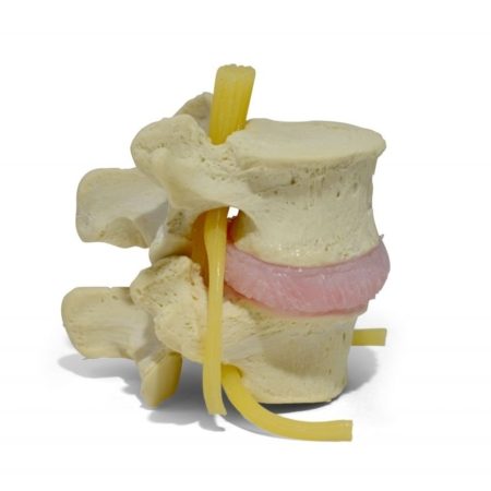
This classic model is a dynamic and constructed to withstand daily clinical use. Take patient education to a dynamic level with an annulus and nucleus.
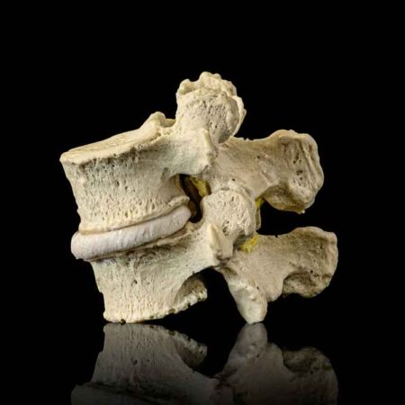
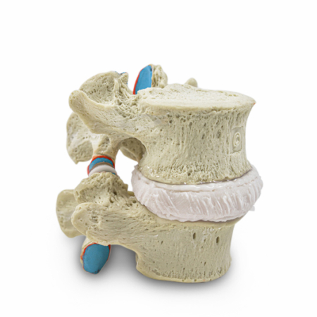
A one-level spine model (L4-5) constructed from a degenerative spine showing disc height loss and facet arthropathy. New updated feature of foraminal stenosis.
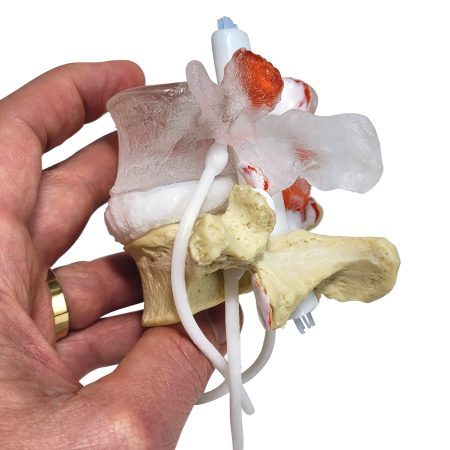
This model has been constructed with a small suction cup and an adjacent polished facet surface to allow the demonstration of an audible release.

Display your model with an acrylic stand. Includes 4 rubber feet and a 1/4 inch round post to allow the model to sit and spin in position.
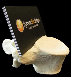
An anatomical way to display your business cards. An L5 vertebra with a notched groove to accommodate standard sized cards.
A lytic pars shows anterolisthesis under load. Designed with an elastomeric two-part disc and the ligamentum flavum to hold posterior elements together.
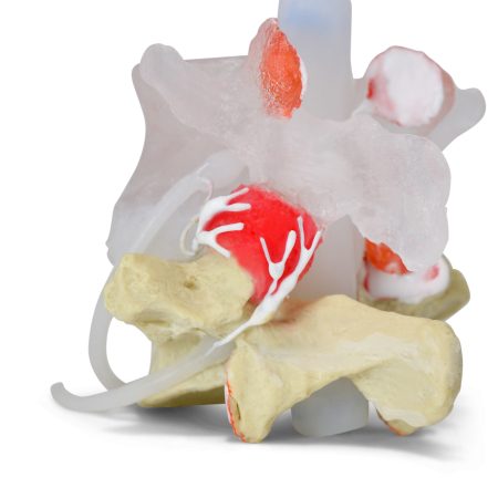
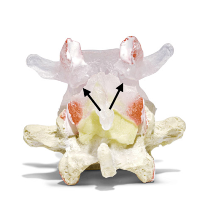
This lumbar model demonstrates unilateral facet innervation branching from three nerve root levels off the posterior primary division.
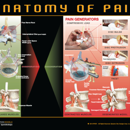
Ad media to your models by incorporating a poster to the wall to drive home the messages related to spine education. More cues to help patients remember.
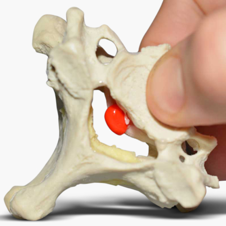
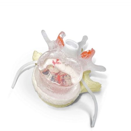
A C6-7 dynamic model showing a central herniating and dynamically extruding nucleus only under load. Show six degrees of freedom with this degenerated model.
Several select models to help educate spine patients with almost any condition. Equip yourself with the ability to teach patients their problems when offering solutions.