
When intervertebral joints bend, they can’t hold as much weight. Limiting lumbar spine flexion may lower the chance of low-back disorders (LBD). However, lifting retraining programs encouraging less lumbar flexion does not always lead to lower LBD rates. This may be because people have trouble keeping their lower backs straight while moving different things. To follow the instruction to keep spine flexion to a minimum, the movement must change to the lower limbs. Let’s say that the range of motion in the lower limb joints is limited or can’t be reached. In that case, people can either bend their lower back more to reach the object or change their stance by rotating their feet outward to lower their spine’s flexion at the start of the lift. Different lift origin heights and masses affect lifting kinematics and kinetics when lumbar spine flexion is limited. It is not well known what these kinematic changes mean for the spine and the joints in the lower limbs. Figuring out these effects could help us determine if lifting retraining programs don’t work because they are too strict or aren’t practical.
To learn more about basic lifting mechanics, researchers can change limits in a planned way and watch how movement coordination changes. Limiting movement options is hard when you try to keep spine bending to a minimum. Other things that limit movement options are lift origin height and mass lifted. Previous research has shown that using a harness to limit the motion of the lower back spine physically lowers lumbosacral compression and increases the range of motion in the lower limb joints compared to a self-selected lifting method. However, these studies only looked at one mass and starting height. When you lift things from lower levels and with heavy loads, your lower limbs and lower back have to move and carry more weight. Changing how biomechanical demands are distributed might also affect other job limitations, such as the amount of time and weight that can be lifted. So, the change in biomechanical demands could be different based on how these constraints interact when the spine is restricted in flexion.
By integrating the power-time curve, you can find the joint work, which measures the biomechanical needs of several joints. Kinematic and kinetic data are part of mechanical energy expenditure (MEE), a complete measure. MEE adds up all the demands across all joints to figure out the total biomechanical demands on the body and how they are distributed when pulling with limited spine flexion.
In 1991, Gagnon and Smyth used MEE to look at joint work at different lift heights and object masses. They found methods that put work on the hips for low lifts and the lower back for higher lifts. But they didn’t look at how mass changes the distribution of MEE at different below-waist heights. This means that it’s not clear how lift origin height and object mass change the distribution of MEE, especially when the spine can’t move as freely.
This research[1.D.R. Carnegie, S.M. Hirsch, T. A. C. Beach, S.J. Howarth, Restricting lumbar spine flexion redistributes and changes total mechanical energy expenditure during lifting, Journal of Biomechanics(2024), doi: https://doi.org/10.1016/j.jbiomech.2024.112132] looks into what happens to total MEE and how it is distributed in the spine and lower limbs when lumbar spine flexion is limited during pulling. The study aims to show that limiting spine flexion will lower MEE in the spine and raise MEE in the lower extremity joints. The highest MEE was seen when heavier items were lifted from lower heights. We also look at how the object’s mass and lift point height change these changes in the MEE distribution.
Twenty participants (10 male, 10 female), aged over 18, were recruited from post-secondary students. Exclusion criteria included current musculoskeletal pain or affirmative responses to the Physical Activity Readiness Questionnaire. Participants provided written informed consent, and the study was approved by the Canadian Memorial Chiropractic College and University of Toronto ethics boards.
Participants wore tight-fitting clothing to facilitate the placement of reflective markers for a 10-camera motion capture system. Marker data were synchronized with two in-ground force platforms.
Participants wore a custom spine harness that restricted lumbar spine flexion. Lift origin heights were set at mid-shank and knee levels, with masses of 20kg and 34kg for females and 34kg and 58kg for males. Eight lifting task combinations were performed, varying spine restriction, lift origin height, and mass. Each task involved five continuous repetitions, lifting a barbell from the platform to an upright posture and returning it.
Marker and force data were processed in Visual3D. Joint angles, moments, and powers were calculated, and MEE for each joint was determined using integrated joint power-time curves. Total and joint-specific MEE values were averaged over five repetitions.
Means, standard deviations, and confidence intervals for MEE and peak joint angles were calculated. Repeated measures analyses of covariance evaluated MEE, with spine restriction, lift origin height, and object mass as factors and lift time as a covariate. Post hoc analyses used Tukey’s adjustment for significant interactions.
The 20 participants had a mean age of 26.0±1.5 years, height of 1.80±0.10 m, and mass of 76.0±14.4 kg. Lift origin heights were 0.42±0.03m (low) and 0.58±0.05m (high), with consistent stance widths and foot orientations.
Total MEE increased with spine restriction, lower lift origin height, and heavier mass. Significant interactions were found between spine restriction, lift origin height, and lift origin height and object mass. Lift time was longer for heavier masses and lower lift origins.
Spine restriction reduced spine MEE and increased hip MEE, particularly for lower lift origins. Lift origin height and mass influenced knee and ankle MEE, with greater MEE for heavier masses at high lift origins and lower lift origins for knees.
Limiting lumbar spine flexion shifts biomechanical duties to the hips. This may explain why lifting retraining programs that don’t look at how these instructions can be carried out don’t work. The change in MEE was affected by the lift origin height but not by the mass of the item. These results show that limiting lumbar spine flexion can shift stress to the lower limbs, but whether or not this is possible and effective depends on the person and the task.
This study highlights the complexity of biomechanical demands in lifting tasks and suggests that directives to minimize spine flexion should account for individual differences and task-specific constraints to be effective.
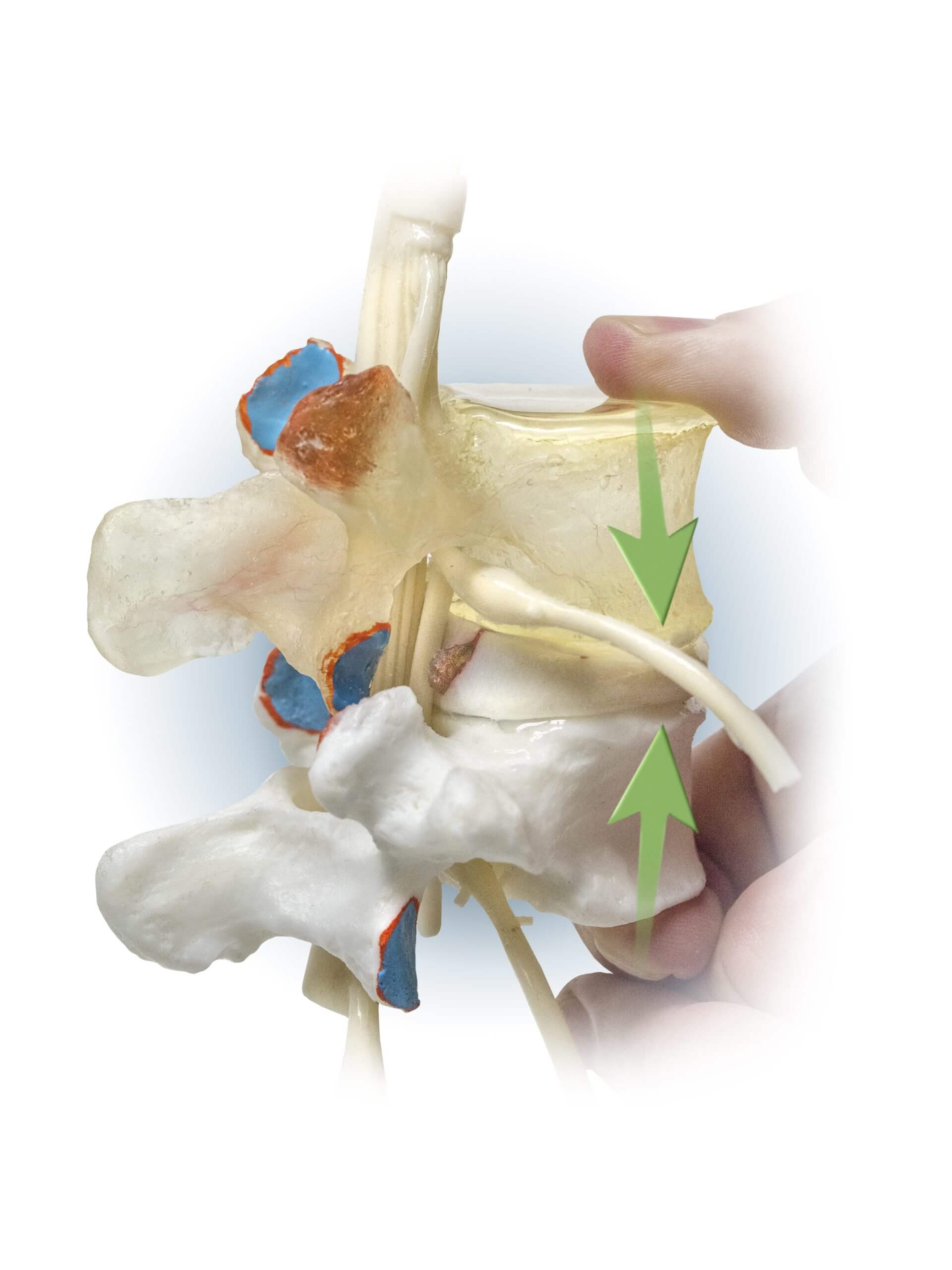
Patient education is critical to preventing low-back disorders (LBD) and teaching proper lifting techniques. A Dynamic Disc Model (DDM) can significantly enhance this educational process. Here’s how:
In summary, using a Dynamic Disc Model for patient education provides a comprehensive, interactive, and visually engaging way to teach proper lifting techniques. By helping patients understand the mechanics of their spine and the impact of their movements, healthcare providers can significantly enhance the effectiveness of their education efforts, ultimately leading to better outcomes in the prevention and management of low-back disorders.
Share Now